Panoramic Radiographic Study on Location of Mental Foramen in Patients of Orthodontic Treatment
Shardha Bai Rathod1, Dr Anand V Nimbal2, Dr Shreedevi K Chillalshetti3 Dr Vinutha Chiniwar4, Dr Vikas Desai5, Dr G A Hadimani6*, Dr.Ishwar Bagoji7 Dr Ambadasu Bharatha8
Lecturer1*, Prof & Head2, Senior Resident3, Senior Resident4, Assistant Professor5,6,7 Department of Dentistry and Anatomy, BLDE (Deemed to be University), Shri BM Patil Medical College, & Research Centre, Vijayapura, Karnataka, India. 8Lecturer in Faculty of Medical Sciences, The University of the West Indies, Cave Hill Campus, Barbados, WI.
Introduction:
The accurate identification of the mental foramen (MF) is important both for the diagnoses and clinical procedures.
The mental foramen is a small foramen situated in the anterolateral aspect of the body of the mandible. The mental foramen is defined as the entire funnel-like opening in the lateral surface of the mandible at the terminus of the mental canal. This foramen is contained entirely within the buccal cortical plate of bone. The most common location of mental foramen is below the apex of the second premolar or between the apices of first and second premolars. The average size of the foramen measured was 4.6 mm horizontally and 3.4 mm vertically on the lateral surface of the mandible. (1, 2)
The accurate knowledge of the position of the mental foramen is important both when administering regional anaesthesia, performing periapical surgery, dental implant surgery, and endodontic treatment. Panoramic radiographic study of Mental Foramen is the most preferred diagnostic modality as it allows more accurate localization of the mandibular foramen in both a horizontal and vertical dimension. (3) The position of the mental foramen varies among races and genders. There have been variations in the mental foramen ranging from the difference in the shape and position to the presence of accessory foramen or even complete absence in some cases. (4) The purpose of this study was to report the usual position of the mental foramen in Vijayapur; it has not been reported in this population before.
Materials and methods
We had evaluated 291 all panoramic radiographs from the patients within the age group of 15-40 years who were advised for OPG for various purposes, most of them were referred for orthodontic treatment to the Dental Department, in Shri B. M. Patil Medical College Vijayapur. Informed consent was taken from all the patients and children's consent parents. The study was approved by the ethical committee. This study was conducted over a period of one year. Radiological evaluation of the patient was done using the OPG model Orthoralix digital panoramic system (Tube Potential: 70-80 kv, Tube Current: 2-15 mA, Total Filtration: >2.5 mm, time: 14.8 s). The magnification factor, as reported by the manufacturer, is 1.25.
Exclusion criteria included:
1) Non-visualization of the mental foramen bilaterally on OPG
2) Presence of impacted premolar
3) Patients with missing teeth and congenitally missing premolar.
4) Presence of a radiolucent lesion in the lower jaw anywhere around mental foramen of the mandible
All radiographs were analyzed by the principal investigator. Random sample of 10 radiographs that were re-examined by the corresponding investigator. The assessment of radiographs was made digitally.
The position of the image of the mental foramen in a panoramic radiograph was recorded according to criteria given by Wei Cheong Ngeow and Yusof Yuzawati.(5,6) They were:
1) Position 1: Situated anterior to the first premolar
2) Position 2: In line with the first premolar
3) Position 3: Between the first and second premolars
4) Position 4: In line with the second premolar
5) Position 5: Between the second premolar and first molar
6) Position 6: In line with the first molar
We used the edge of a ruler for the identification of the longitudinal axis of the nearest tooth on both the side and the position of the mental foramen was recorded with respect to that. The position of the large mental foramen situated between two teeth was established after drawing an imaginary line in parallel to the long axis of the teeth.[6] The location of the mental foramen was reported on the basis of gender and symmetry or asymmetry.
Statistical analysis
For anticipated Proportion of common position of the mental foramen between first and the second premolar 71.5% the minimum sample size is 313 patients with a 5% level of significance and 5% absolute error. Data was represented by using Mean+/-SD, percentages and diagrams. And a significant difference was found using the chi-square test. Study duration: 1 year
The formula used: n=z2 p*q / d2
Results
Of the 291 panoramic radiographs of patients was analyzed, 83 were that of males and 208 that of females. The female patients were more compared to male patients who had been referred for the aesthetic process of orthodontic treatment. [Table 1 &2]
TABLE: 1 DISTRIBUTION OF CASES ACCORDING TO AGE AND GENDER
|
Age (Yrs.) |
Male |
Female |
p-value |
||
|
Frequency |
% |
Frequency |
% |
||
|
15-20 |
58 |
28.7 |
144 |
71.3 |
0.94 |
|
20-25 |
20 |
27.0 |
54 |
73.0 |
|
|
25-30 |
3 |
37.5 |
5 |
62.5 |
|
|
>40 |
2 |
28.6 |
5 |
71.4 |
|
|
Mean age |
19+/-9.3 |
19+/-6.7 |
|||
|
Total |
83 |
28.5 |
208 |
71.5 |
|
A total of 291 patient's OPGs were examined in the study. Of that 208 were females (71.5%) and 83 were males (28.5%). The most common position for the mental foramen in the present study sample was position 4 (54.2%) followed by position 3(37.5%) [Table 3]. A total of 116 0n left(40%) and 291 on right (63.1%) were found in position 4 inline second premolar and a total of 140 on left(48.3%) and 142 on right (30.8%) were found in position 3, between first and second premolar.
TABLE: 2 GENDER WISE DISTRIBUTION OF MENTAL FORAMEN
|
LOCATION |
MALE |
FEMALE |
TOTAL |
|||||||||
|
LEFT |
RIGHT |
LEFT |
RIGHT |
LEFT |
RIGHT |
|||||||
|
N |
% |
N |
% |
N |
% |
N |
% |
N |
% |
N |
% |
|
|
1 |
0 |
0.0 |
0 |
0.0 |
0 |
0.0 |
0 |
0.0 |
0 |
0.0 |
0 |
0.0 |
|
2 |
1 |
1.2 |
1 |
0.7 |
7 |
3.4 |
8 |
2.5 |
8 |
2.8 |
9 |
2.0 |
|
3 |
40 |
48.8 |
39 |
28.9 |
100 |
48.1 |
103 |
31.6 |
140 |
48.3 |
142 |
30.8 |
|
4 |
29 |
35.4 |
83 |
61.5 |
87 |
41.8 |
208 |
63.8 |
116 |
40.0 |
291 |
63.1 |
|
5 |
12 |
14.6 |
12 |
8.9 |
14 |
6.7 |
7 |
2.1 |
26 |
9.0 |
19 |
4.1 |
|
6 |
0 |
0.0 |
0 |
0.0 |
0 |
0.0 |
0 |
0.0 |
0 |
0.0 |
0 |
0.0 |
|
TOTAL |
82 |
100 |
135 |
100 |
208 |
100 |
326 |
100 |
290 |
100 |
461 |
100 |
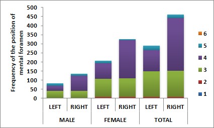
FIGURE:
GENDER WISE DISTRIBUTION OF MENTAL FORAMEN
The mental foramina are usually found to be symmetrically located on both the side.
TABLE:3 ASSOCIATION OF ASYMMETRY/SYMMETRY AND GENDER
|
ASYMMETRY/SYMMETRY |
MALE |
FEMALE |
p-value |
||
|
N |
% |
N |
% |
||
|
Asymmetry |
33 |
39.8 |
56 |
26.9 |
0.032* |
|
Symmetry |
50 |
60.2 |
152 |
73.1 |
|
|
Total |
83 |
100 |
208 |
100 |
|
Note: * significant at 5% level of significance (p<0.05)
Discussion
The mental foramen (MF) is a very important landmark during surgical procedures. To avoid mental nerve injury during surgical procedures and an elongated styloid process leads to inferior alveolar nerve irritation during mandibular movement. This results in a progressive loss of sensation in the mental nerve. (7) The knowledge of the site of the mental foramen in the patients allows for accurate delivery of local anaesthesia for dental procedures and avoids damage to the nerve in surgical procedures, such as periapical surgery, implantation, cyst enucleation, periodontal surgery, mandibular bony osteotomy. (8)
Additionally, good interpretation of anatomical landmarks in oral pathology and forensics can be aided. For all of the above-mentioned reasons, the horizontal location and position of the mental foramen have been studied utilizing panoramic radiographs or dry skulls.
With respect to detecting the anatomical structure, the cadaveric dissection methods are more accurate than radiographic methods. It has been suggested that the most common position of MF examined on 525 dry mandibles was in 43.66% located in front of the apex of the root of the second premolar that was position 4. (9)
A different position of mental foramen has recently been reported to have existed between the population of different or even of the same geography. (10) In this study, the position of the mental foramen in a randomly selected patient in the Vijayapur population was studied using panoramic radiographs rather than other plane films. Consequently, a more accurate interpretation of the location of the mental foramen in both the horizontal and vertical dimensions was allowed by using panoramic radiographs. (11, 12)
In our series of 291 panoramic radiographs, the position of mental foramen was most commonly both in males and females in line with the second premolar and the second most commonly was between first and second premolar. No cases were found in positions 1 and 6. This present study result coincides with observations made by Ngeow et al. The previous study showed that 95% of mental foramen was reported the position of the 3 and 4. Studies done on North American and Caucasian populations reported the area between the two premolars as the most common location of the mental foramen. (13, 14, 15) Our study on the position of mental foramen was inconsistent with findings of Yosue and Brooks,& Gupta and Soni.(12,1) The Panoramic study of position of mental foramen was also observed in Tanzanian adult black male(16), Malay population(13) Brazilian,(17) Sankar et al.,(18) Malawian mandible,(19) Ukoha (20) in southeastern Nigerian Singh et al. (21) and Kanta et al. Jasser and Nwoku observed most common position as in line with the longitudinal axis of second premolar followed with location between first and second premolar in radiographs study of Saudi Arabians. (22)
We had selected patients with age group from 15 to 40, the present study is a cross-sectional, epidemiologic convenient study, and was done in BLDE Medical Hospital in Dental Department, a period of one year. The presence of premolar for the detection of the mental foramen is important, from the age of 11years the lower canine and premolar starts erupting, so we selected the patients from the age group of 15 years and above. The study excluded patients with impacted premolar, non erupted premolar, and congenitally missing premolar, fractured cases of mandibular in region and radiograph defects.
We used panoramic radiographs because, the wide area of both hard and soft tissues is visualized in continuity, thus allowing a more accurate location of the mental foramen in both horizontal and vertical dimensions. The research results show different variability and asymmetry in the position and shape of the mental foramen in different races, age, sexes and geographies.
Conclusion
Accurate knowledge is important for dental practitioners about variation in anatomical and morphological appearance and position of the Mental Foramen for the isolation of mental nerves and vessels is to perform safer mental nerve blocks in surgical interventions.
Acknowledgment: I would like to thank Dr. Anand V Nimbal corresponding investigator and HOD of the Dental Department of BLDE (Deemed to be University) hospital Vijayapur for helping me in the observation of the panoramic radiographs.
References:
[1] Gupta S, Soni JS. Study of anatomical variations and incidence of mental foramen and accessory mental foramen in dry human mandibles. Natl J Med Res 2012;2:28-30. [Google Scholar]
[2] Muhammad DA. Anteroposterior position of the mental foramen on panoramic radiographs in Sulaimani population. Kurdistan Acad J 2009;8:9-16. [Google Scholar]
[3] Gungor K, Ozhurk M, Semiz M, Brooks SL. A radiographic study of location of mental foramen in a selected Tuekish population on panoramic radiograph. Coll Antropol 2006;30(4):801. [Google Scholar]
[4] Hasan T, Fauzi M, Hasan D. Bilateral absence of mental foramen - A rare variation. Int J Anat Variations 2010;3:167. [Google Scholar]
[5] Haghaifar S, Rokouei M, Radiographic evaluation of the mental foramen in a north American, , population w. . 1998;85:457-460. [Google Scholar]
[6] Ngeow WC, Yuzawati Y. The location of the mental foramen in a selected Malay population. Journal of Oral Science 2003;45(3):171-175. Available from: http://joi.jlc.jst.go.jp/JST.Journalarchive/josnusd1998/45.171?from=CrossRef doi: 10.2334/josnusd.45.171. [Google Scholar]
[7] Bagoji IB, Hadimani GA, Patil BG, Bannur BM, Ambadasu B. Bilateral elongated styloid process: Its anatomical embryological and clinical implications. International Journal of Medical Research & Health Sciences 2013;2(2):273-6. Available from: http://www.indianjournals.com/ijor.aspx?target=ijor:ijmrhs&volume=2&issue=2&article=026 doi: 10.5958/j.2319-5886.2.2.015. [Google Scholar]
[8] Rowe AH. Damage to the inferior dental nerve during or following endodontic treatment. Br Dent J 1983 Nov;155(9):306-7. Available from: http://www.nature.com/articles/4805219 doi: 10.1038/sj.bdj.4805219. [Google Scholar]
[9] Gershenson A, Nathan H, Luchansky E. Mental foramen and mental nerve: changes with age. Acta Anat (Basel 1986;126(1):21-8. Available from: https://www.karger.com/Article/FullText/146181 doi: 10.1159/000146181. [Google Scholar]
[10] Ari I, Kafa IM, Basar Z, Kurt MA. The localization and anthropometry of mental foramen on late Byzantine mandibles. Coll Antropol 2005;29(1):233-6. [Google Scholar]
[11] Philips JL, Weller, R. Norman, , Kulild JC. The mental foramen: Part II. Radiographic position in relation to the mandibular second premolar. Journal of Endodontics 1992;18(6):271-274. Available from: https://linkinghub.elsevier.com/retrieve/pii/S0099239906809532 doi: 10.1016/S0099-2399(06)80953-2. [Google Scholar]
[12] Yosue T, Brooks SL. The appearance of mental foramina on panoramic radiographs. I. Evaluation of patients. Oral Surgery, Oral Medicine, Oral Pathology 1989;68(3):360-364. Available from: https://linkinghub.elsevier.com/retrieve/pii/0030422089902247 doi: 10.1016/0030-4220(89)90224-7. [Google Scholar]
[13] Ngeow WC, Yuzawati Y. The location of the mental foramen in a selected Malay population. Journal of Oral Science 2003;45(3):171-175. Available from: http://joi.jlc.jst.go.jp/JST.Journalarchive/josnusd1998/45.171?from=CrossRef doi: 10.2334/josnusd.45.171. [Google Scholar]
[14] Shankland WE. The position of mental foramen in Asian Indians. J Oral Implantol 1994;68:118-23. [Google Scholar]
[15] Gangotri S, Patni VM, Sathwane RS. Radiographic determination of position and symmetry of mental foramen in Central Indian population. J Indian Acad Oral Med Radiol 2011;23:101-3. Available from: http://www.jaypeejournals.com/eJournals/ShowText.aspx?ID=1357&Type=FREE&TYP=TOP&IN=~/eJournals/images/JPLOGO.gif&IID=115&isPDF=YES doi: 10.5005/jp-journals-10011-1104. [Google Scholar]
[16] Fabian FM. Position, shape and direction of opening of the mental foramen in dry mandibles of Tanzanian adult black males. Ital J Anat Embryol 2007;112:169-77. [Google Scholar]
[17] Maise MA, Felippe BP. The mental foramen position in dentate and edentulous Brazilian's mandible. Int J Morphol 2008;26:981. [Google Scholar]
[18] Sankar DK, Bhanu SP, Susan PJ. Morphometrical and morphological study of mental foramen in dry dentulous mandibles of South Andhra population of India. Indian J Dent Res 2011;22(4):542-6. Available from: http://www.ijdr.in/text.asp?2011/22/4/542/90290 doi: 10.4103/0970-9290.90290. [Google Scholar]
[19] Igbigbi PS, Lebona S. The position and dimensions of the mental foramen in adult Malawian mandibles. West Afr J Med 2005;24:184-9. [Google Scholar]
[20] Ukoha UU. Position, shape and direction of the mental foramen in mandibles South-Eastern Nigeria. Int J Biomed Res 2013;4(9):499-503. Available from: http://ssjournals.com/index.php/ijbr/article/view/876 doi: 10.7439/ijbr.v4i9.349. [Google Scholar]
[21] Roy PP, Ambali MP, Doshi MA, Jadhav SD. Variation in the position shape and direction of mental foramen in dry mandible. Int J Anat Res 2014;2(2):418-20. [Google Scholar]
[22] Jasser NM, Nwoku AL. Radiographic study of the mental foramen in a selected Saudi population. Dentomaxillofac Radiol 1998;27(6):341-3. Available from: http://www.nature.com/doifinder/10.1038/sj.dmfr.4600388 doi: 10.1038/sj.dmfr.4600388. [Google Scholar]